Video - Magma Arta: Rocks under the Microscope, Sedgwick Museum, Cambridge University
http://www.microscopy-uk.org.uk/mag/artfeb04/iwouslides2.html
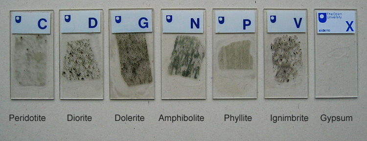
I am fortunate in that my husband owned the materials and microscope relating to this OU course.
I used the microscope to create micro paintings which I spent hours viewing and manipulating with an etching needle and experimenting with ink, paint and varnish. See post: Transformers.
Selected slides for the image gallery.
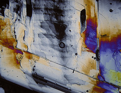
General: Gypsum is a mineral and appears colourless in thin sections, it is the chief constituent of gypsum rock. Gypsum when accurately cut to the required thickness was commonly used with polarizing microscopes as a 'sensitive tint' plates or 1st order red plates which could be inserted into the light path of a microscope using crossed-polars.
This slide: Under plane polarised light you can clearly see one of the cleavage planes and from the crossed-polar image some of the interference colours. Gypsum has a low birefringence value of 0.009 and would normally show only weak interference colours of the 1st order similar to quartz in thin section, but this section is much thicker in parts and showing stronger interference colours.
Slide [C], Peridotite from Italy.
General: Peridotite is an igneous rock comprising mostly olivine, pyroxene and hornblende. Chromite [a source of chromium] is often associated with peridotite.
  Some images inspire painting while others inspire drawing |
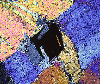
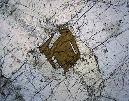
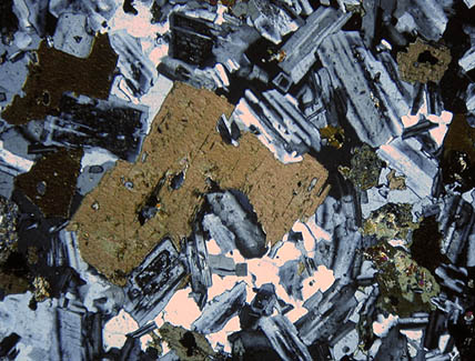
General: Diorite is an igneous rock.
This sample: the left and right images are from different parts of the slide, in the crossed-polar image a central crystal thought to be biotite can be seen surrounded by feldspar showing various degrees of twinning.
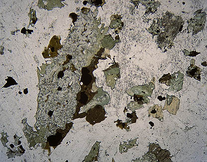 Slide [D], Diorite from Italy. |
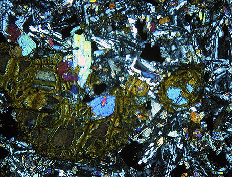
Slide [G], Dolerite from Clee Hill.
General: Dolerite is an igneous rock and is one of the varieties of basalt and coarse grained in texture.
|
No comments:
Post a Comment